Preventing breast cancer is possible. The answer is under your nose & right in your city,
Check breasts annually with breast thermography!
but your Medical Doctor is likely not telling you about it because Mammograms are the fad and have been for many years…but unfortunately they are not preventative because they show calcifications/small lumps etc…abnormalities that are already present. And they are not perfect, they can miss finding a lump. Not too mention if you do have a lump it can squish it and possibly leak out cancer cells into your lymphatic system and lastly the radiation though small, is still radiation.
If I told you that there was a method that has been around for years and can 90% of the time find pre-cancerous conditions in your breasts, and that if found there is an actual dietary protocol & herbs to help get rid of the cancerous conditions and get your breast back to normal….what would you say??
Breast Thermography, otherwise known as Digital Infrared Imaging, is what it is called and has been done by professionals for years, but not in your hospital, in private offices across Canada. They are read by a Radiologist and your breasts are rated from a TH-1 to a TH-5, with 1 being the most normal and 5 being the most abnormal. The cost is $250.00 and can be claimed under some benefits plans as a “laboratory test”.
My purpose here is to show the differences between the diagnostic tests out there for breast cancer and show women why breast thermography is preventative compared to the other methods, that do also have there place.
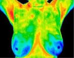
Digital Infrared Imaging
Digital Infrared Imaging scans using a digital infrared imaging camera and a high speed computer and shows the function, the physiology, the metabolism of breast tissue. It gives a picture of the functional activity in the breast tissue. Usually abnormal cells are hotter because a malignant tissue mass is greedy. To feed their rapid growth they produce a chemical that makes new blood vessels grow, this is called angiogenesis (“angio” means “blood vessel”, “genesis” means “creation”.). A digital infrared imaging scan shows the heat difference between normal breast tissue and problem areas. In scientific terms, the normal breast tissue acts as the control against which any hot areas are compared.
Not all malignancies are hyper-vascular, that is, a small number do not show increased blood supply. Unless there are other signs, a thermography scan will not detect a non-hypervascular malignancy.
Which means you still need to feel your breasts monthly to make sure you do not feel any lumps since if there is a lump you would want to get other diagnostic tests done besides a thermography.
A Digital Infrared Imaging Scan provides the earliest evidence of breast disease. The sensitivity rate is 90%. This means in 90% of the cases, the scan accurately indicates a presence or absence of disease. Digital infrared imaging has a 10% false positive rate; in 10% of the cases the results may suggest disease where there is none.
Ultrasound
Ultrasound uses sound waves aimed at breast tissue. The sound echoes back when it encounters a mass. Sent to a computer, the echo waves show the shape and density of the mass. When strict criteria are used, ultrasound can distinguish between a simple benign cyst and other masses.
A negative ultrasound does not mean there is not a breast mass. There may be a mass, but if it has the same density as other breast tissue, no echo is produced.
Ultrasound is not a screening method. It is a follow-up diagnostic tool to explore the shape and edges of a breast mass which suggest whether the mass is benign or malignant.
Magnetic Resonance Imaging (MRI)
Like ultrasound, MRI is not a screening method but a follow-up diagnostic tool, MRI is being studied for its ability to distinguish between benign and malignant tissue masses, and to help physicians and the patient to decide whether a lumpectomy or mastectomy should be done. MRI’s area also used to examine breast implants to see if they are leaking. While ultrasound and mammography can only visualize the shell of the breast implant, MRI can tell if there is leakage.
MRI is not a screening method. It is a follow-up diagnostic tool useful for breast implants. Its role in breast disease and breast cancer is still being studied.
Mammography
Mammograms show the physical anatomy or structure of the breast. When an x-ray encounters a mass, it does not pass through as easily as it does through normal breast tissue so the mass shows up as a white area on the x-ray. Medical guidelines recommend a baseline mammogram so physicians can compare the first one with later ones. When a mass is found, an ultrasound may be done to get more information about the size and shape because the mass could be a dense cyst. At this stage a needle biopsy may be recommended.
Mammograms work best for soft post-menopausal breast tissue, and for slow-growing tumours. Mammograms do not view the whole chest wall; they are not effective for young, dense breast tissue, large breast, fibrocystic breasts, enhanced breasts (implants) and breast of women who are pregnant, breastfeeding, or on hormone replacement therapy (HRT).
Mammograms are used for breast cancer screening and show the location of a mass or tumour. Mammograms have a 25% false positive rate, meaning that 25% of the cases results suggest disease where there is none. They have a 20% false negative rate (missed tumours) mostly in younger women, but can be missed in older women as well, due to the density of the breast tissue of women under 50 years of age.
A breast biopsy provides the only definitive diagnosis of breast cancer. All other methods are investigative and adjunctive diagnostic tools.
So what is the big deal?? Well, here are the benefits of using the Digital Infrared Imaging Screening:
- It is timely. Problems can be found before abnormalities are seen with mammograms. Early detection provides the best outcome and treatment can be provided to actually improve your breast rating. Normally, it is a diet & supplements that actually will remove the offending substances from the breast tissue Ie. Xenoestrogens. (False-estrogens from plastics, pesticides etc.)
- Inclusive. Which means that it examines the whole chest, breasts and armpit areas.
- Good for all ages and stages. It is good for puberty, pregnant, breast-feeding, pre-menopausal years & post-menopausal years.
- Good for all breast types. Good for dense, pregnant, breastfeeding, fibrocystic, enhanced (implants) and women on oral hormone medication either BCP, HRT or Bio-identical Hormones.
- Painless. No squeezing, no pressure, no touching by equipment or technician.
- Risk-free. No harmful rays emitted so digital infrared imaging scan can be done as often as needed to monitor breast health and to guide treatment.
- Risk indicator. Digital infrared imaging results are a better indicator of future breast disease, than a family history of disease.
So what happens in a Digital Infrared Imaging Screening Session?
Preparation Stage (about 20 minutes)
First, you will be asked to fill out a breast history form about symptoms related to possible breast dysfunction and disease. In a private room, you will undress to the waist and let your breasts adjust to the cool room temperature (18-22 C). It takes about 10 minutes for this part so you can relax or read.
Screening Stage (about 10 minutes)
You will be asked to stand with your hands on your head about 10 feet in front of a digital infrared imaging camera. there images will be taken: straight-on, and right and left partial side views.
Cold Challenge
You will be asked to put both hands in cool water (about 10 C) for 1 minute. This is a cold challenge to your blood vessels. Normal blood vessels narrow and gradually become cooler with this challenge while abnormal vessels do not narrow and remain warmer.
After the cold challenge, a second series of three scans will be taken to record the changes in the response of the blood vessels to the cold challenge.
Report Stage
Your digital infrared imaging breast scans will be read and analyzed by a member of the American Board of Thermology. You will receive your report by email. There will be given and estimated expected date the report will be sent to you. It is also recommended that you send a copy of your report to your N.D., M.D., and any other health care providers.
Please note: this technology can be used for other parts of the body, Ie. Thyroid. Always inquire if you feel you want another part of your body scanned.
Thermology Report
The thermology report, written in precise technical language, is summarized by a number which is a classification guide regarding breast tissue function. The criteria for breast thermology classifications were established in 1970 at the Pasteur Centre in Paris. These classifications, known as the marseille system, are reported on a scale ranging form TH-1 to Th-5.
The numbers are an indication of normal (TH-1) to very abnormal (TH-5) cell function. The higher numbers indicate a higher risk of malignant disease, but THEY ARE NOT A DIAGNOSIS OF CANCER. The only accepted diagnosis of breast cancer is form a microscopic examination of cells from a biopsy.
All reports should be discussed with your health care professional. Your health care professional will determine what follow-up testing and treatment is required. The report will suggest or recommend a re-test timeframe.
All reports should be discussed with your health care professional. Your health care professional will determine what follow-up testing and treatment is required. The report will suggest or recommend retest timeframe.
TH-1
Is normal, healthy breast tissue: no evident, suspicious hot spots showing increased metabolic activity. Next screen: usually 1 year.
TH-2
Normal, uniform breast tissue: some blood vessel activity (hotter than TH-1, but responds normally to cold challenge). Benign cysts and elevated estrogen levels are in this category. Next screen: usually 1 year. Using a dietary protocol with a supplement protocol would be useful in cleaning up the breast tissue to see if you can get to the lowest level TH-1.
TH-3
Suspicious cell activity: higher heat areas in breast tissue. Next screen: Usually within 6 months to monitor. And I would do a dietary and supplement protocol to help clean up the breast tissue, this way you increase your chances to have a normal thermography the next time.
TH-4
Abnormal cell activity: higher heat areas in breast tissue. A dietary & supplement protocol is required to clean up the breast tissue and bring it back to normal. Next screen: Usually within 3 months to monitor.
TH-5
Severely abnormal cell activity: higher heat areas in breast tissue. Again, a dietary & supplement protocol to clean up the breast tissue is required to bring the breast tissue back to normal. Next Screen: Usually within 3 months to monitor
Digital Infrared Imaging Practitioner:
Suzanne Sutherland, Certified Thermographic Technician. She has an office both in Vancouver and Calgary. She is very professional and would highly recommend her. Call or email to book your appointment. She is only in Calgary every 3 months so book your appointment today!
CALGARY
26 Discovery Hts, SW
Calgary, AB T3H 4Y6 Phone number: (778) 340-1947
http://www.breastthermography.ca/
Short Case Review:
50 year old caucasian women with strange breast pain/sensations and have persisted for 3 months. There were no lumps found when she palpated her breasts. She went for a breast thermography and the right breast was TH-4 and the left breast was TH-5.
For 6 months she did the Gerson Cancer Therapy diet and took lymphatic herbs particular for breast tissue and her constitutional type. She repeated the breast thermography and her breast tissue had gone down to TH-1 & TH-2.
Her breast sensations were also gone and there were still no lumps detected by palpation. In this case, as you can see, breast thermography helped to show the abnormal changes prior to developing breast cancer so that she was able to take a course of action to change the tissue back to normal.
I believe this can happen for more women!! We do not have to have our breasts removed. I do not understand the politics of why thermographies are not done in the hospitals, but I can tell you that cancer brings in a large amount of money and how much money would be lost in breast cancer treatment if breast cancer was never diagnosed in the first place?? Thousands of dollars.
Make breast thermography an annual part of your health program. Don’t wait.
Start today!
Note: this information is provided for information purposes only and is not to diagnose or treat anyone.
 In the past few years we’ve heard a lot of talk and read numerous books about the relationship between inflammation and chronic disease. We now know, for example, that inflammation is at the root of heart disease, cancer, autoimmune disorders, asthma, allergies, arthritis, and deteriorating mental ability in aging.

 Chemical damage comes from toxins and toxins derive from three main sources. First, there are poisons which can be introduced into the body. These could be plant toxins (poison ivy, death cap mushrooms, etc.) or animal venoms (bee stings, spider bites, etc.) or chemicals (heavy metals, gasoline, solvents, etc.). Second, there are metabolic toxins (waste products of our own metabolism. And third, there are metabolic toxins released by microorganisms and parasites (such as yeast, bacteria, intestinal worms, etc.).
 At this stage, inflammation is actually quite easy to reverse. If one can keep the fluid moving out of the tissues and into the lymphatic system, then the cells never suffer oxygen and nutrient deprivation or a localized build up of toxins. This allows repair to take place very rapidly. All one has to do is keep the lymphatics moving, which can be done by applying pressure to the injury, lightly rubbing or massaging the injured area or even applying energy to the area of injury.